
Accueil > Équipes scientifiques > Nanomédecine et Biophotonique (NanoBio) > Imagerie de fluorescence à l’échelle nanoscopique pour la biologie > Microscopies au delà de la limite de diffraction > Microscopie de fluorescence supercritique (SAF) > Supercritical angle fluorescence microscopy (SAF)
Supercritical angle fluorescence microscopy (SAF)
par - 3 août 2015
-Supercritical Angle Fluorescence Microscopy - SAF
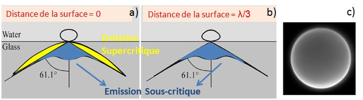
Fluorophores emission directions dependent on their distance to the surface of the glass slide (see Fig.). The fluorophores are similar to nano-antennas with evanescent electromagnetic components when their immediate environment is homogeneous. By cons, when an interface is present at less than a few tens of nanometers, a portion of these components becomes propagating. This signal is then transmitted into the glass in directions beyond the critical angle. Thus, while the subcritical emission is identical whatever the distance fluorophore / glass, only the fluorophores located in the immediate vicinity of the interface have such supercritical emission.
This light sometimes referred to as "forbidden light" may represent up to 50% transmission to the lamella of the fluorescent emitter.
This supercritical emission decreases very rapidly with the distance to the surface and provides an absolute location of nanometric axial fluorophores.
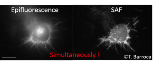
Our work on the development of microscopes field associated with high numerical aperture objective to collect the supercritical issuance. Various devices for isolating and / or format supercritical components has been made (T. Barroca Thesis (ESPCI Langevin Institute)) to detect simultaneously the epi-fluorescence image and the membrane image. The association with the lateral super-resolution techniques like STED and SMLM are ongoing.
SAF Microscopy is an ideal tool also in conventional wide field microscopy as it provides an original tool to access the real-time observation of membrane and adhesion process.
It offers many advantages compared to technical currently offered commercially, particularly microscopy techniques TIRF (Total Internal Reflection Fluorescence). It is based on a complementary approach, the spatial selectivity of not being derived thanks to a confined excitation but by a selectivity at the collection of the sample fluorescence emission. This has many advantages including : significantly background reduction due to the intrinsic light scattering in biological media, to overcome the difficulties of lighting (which provides a more uniform field of view and a cost reduced implementation), and finally it allows to acquire simultaneously and in parallel a standard epi-fluorescence image. This technique can also be coupled with most existing microscopy techniques.
Dans la même rubrique :